




Phage Therapy for Epidermolysis Bullosa: Australian Consortium Targets Chronic Wound Infections With Topical Bacteriophages
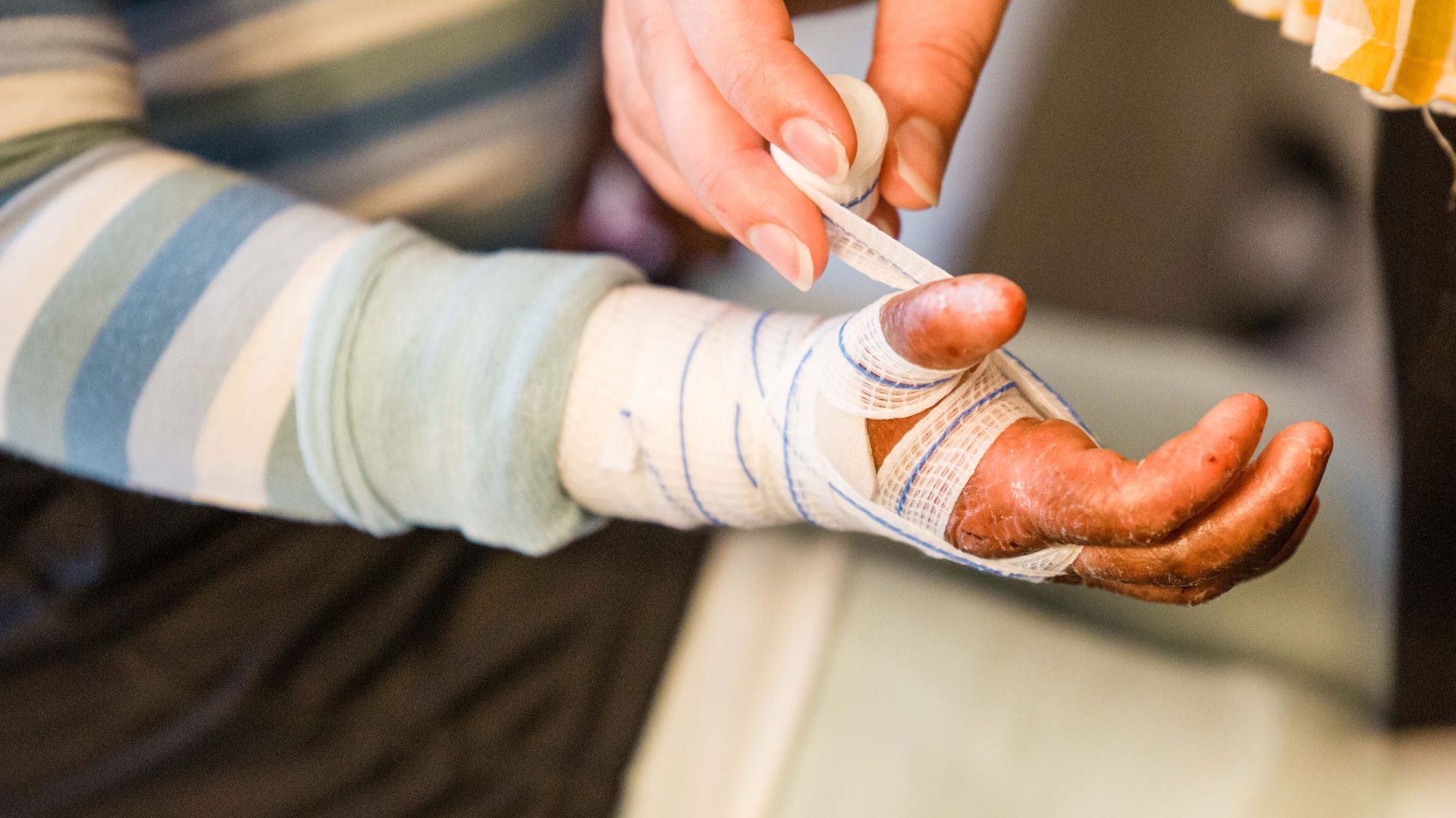
Epidermolysis bullosa is a group of rare inherited disorders characterized by extreme fragility of the skin and, in some forms, the mucosal surfaces. In affected individuals, minor friction or trauma can cause blistering, tissue separation and painful wounds that may remain open for prolonged periods. What begins as a genetic defect affecting the structural integrity of the skin can therefore evolve into a lifelong clinical burden involving chronic inflammation, repeated dressings, scarring, impaired mobility and recurrent bacterial infection. For patients and families, wound care is not an occasional medical intervention but a continuous part of daily life. Against this background, a new Australian research collaboration is investigating whether bacteriophages can be developed as topical treatments to control the bacteria colonizing epidermolysis bullosa wounds while reducing dependence on antibiotics.

The project brings together Adelaide University, AusHealth, EB Research Partnership Australia and the SMART Cooperative Research Centre. Announced in July 2026, it represents the first formal collaborative project supported through SMART CRC and involves a joint investment exceeding one million Australian dollars over two and a half years. The research will be conducted in South Australia under the leadership of Professor Sarah Vreugde at the AusHealth Phage Therapy Centre in Adelaide. Rather than beginning with an established medicinal product, the programme will identify, purify and combine bacteriophages capable of targeting bacterial species commonly found in the chronic wounds of people living with epidermolysis bullosa. The resulting preparations are intended for local application directly to the wound surface, creating a development pathway for a treatment that could eventually help prevent infection, support bacterial clearance and improve wound healing.
The scientific rationale is closely connected to the biology of epidermolysis bullosa. Healthy skin provides a highly effective physical and immunological barrier against microorganisms. In epidermolysis bullosa, repeated blistering and erosion disrupt that barrier, creating exposed tissue surfaces that can be colonized by bacteria from the skin, the surrounding environment and healthcare settings. Chronic wounds can provide nutrients, moisture and protected microenvironments that permit bacterial persistence. Over time, these organisms may organize into biofilms: structured communities surrounded by a self-produced matrix that attaches them to damaged tissue and reduces their susceptibility to antibiotics, antiseptics and host immune responses. A wound can consequently remain colonized even after repeated treatment, while episodes of local infection may delay closure, increase pain and contribute to further tissue damage.
The distinction between bacterial colonization and overt infection is especially important in chronic wounds. The presence of bacteria does not necessarily mean that every wound requires systemic antimicrobial treatment, but high bacterial burden, pathogenic species, virulence factors and biofilm formation can collectively interfere with tissue repair and increase the risk of invasive infection. In patients whose skin is repeatedly damaged, opportunities for recolonization remain constant. Conventional antibiotics may temporarily suppress susceptible bacteria, yet repeated courses can select for resistant populations and disturb the surrounding microbial community. Some drugs also penetrate biofilms poorly, while bacteria in slow-growing physiological states may become less vulnerable to antibiotics that act most effectively during active replication.
Bacteriophages offer a different biological mechanism. They are viruses that recognize susceptible bacterial cells, inject their genetic material and use bacterial machinery to produce new viral particles. Lytic phages ultimately rupture the infected bacterium, releasing progeny capable of infecting additional cells. Their activity is generally much more specific than that of conventional broad-spectrum antibiotics, meaning that a therapeutic phage can attack its bacterial target without necessarily eliminating large fractions of the surrounding microbiota. This specificity is particularly attractive for chronic skin conditions, where the objective is not to sterilize the entire wound ecosystem but to reduce clinically harmful organisms while preserving as much normal microbial balance as possible.
The Australian programme will focus on identifying combinations of phages active against several bacterial types commonly associated with epidermolysis bullosa wounds. This need for combinations reflects a central challenge in therapeutic phage development. Individual phages often infect only a subset of strains within a bacterial species. A virus active against one clinical isolate may be ineffective against another because the bacterial surface receptor is absent, masked or modified, or because intracellular defence systems prevent productive infection. A phage cocktail can broaden bacterial coverage by combining viruses with complementary host ranges and, ideally, different receptor dependencies. It may also reduce the probability that a single resistance mechanism will protect the entire bacterial population.
Developing such a cocktail requires much more than collecting viruses that produce visible zones of lysis in a laboratory assay. Candidate phages must be characterized genomically to exclude undesirable genes, including lysogeny-associated functions, known toxins or antimicrobial-resistance determinants. Their activity must be tested against a representative collection of bacterial isolates recovered from patients rather than against only one laboratory strain. Researchers must evaluate the stability of each phage, its capacity to remain infectious under conditions relevant to the skin and wound environment, its compatibility with other members of the cocktail and its ability to withstand purification, storage and formulation. A preparation intended for damaged human tissue must also be sufficiently pure, particularly with respect to bacterial debris, endotoxins and other contaminants generated during phage production.
Topical administration provides several potential advantages. Applying phages directly to the wound can create a high local concentration at the site where bacteria are growing, without requiring the viruses to survive passage through the gastrointestinal tract or circulate through the bloodstream. Local treatment may also limit systemic exposure and make repeated administration easier. However, wounds are biologically complex environments. Proteins, extracellular DNA, necrotic tissue, dressings, antiseptic residues, pH variation and biofilm matrices can all affect phage diffusion and infectivity. A topical product must therefore be designed not only around the bacterium and the phage, but also around the physical conditions present on the wound surface.
The future formulation could take several forms, although the project announcement does not yet specify the final product design. Phages might ultimately be delivered as a liquid, gel, impregnated dressing or another local formulation capable of maintaining viral activity while providing sustained contact with the wound. Each option presents different manufacturing and clinical questions. A liquid may distribute easily but be removed rapidly by exudate or dressing changes. A gel may improve retention but must not inactivate the phages. A phage-containing dressing could offer prolonged exposure but would require careful control of release kinetics and storage stability. Determining how bacteriophages interact with existing wound-care materials will therefore be an important aspect of eventual translation.
The project is also notable because it is being developed around a rare disease community rather than treating epidermolysis bullosa as a secondary application of a more general antimicrobial platform. EB Research Partnership Australia brings the perspective of patients and families directly into the research structure. Its chair, Nathan Burmeister, described the collaboration as a new model of patient-led research in which community-raised funds are multiplied through institutional partnerships. This involvement can influence which outcomes are considered genuinely meaningful. Laboratory reductions in bacterial counts are important, but patients may prioritize changes in pain, odour, exudate, dressing frequency, wound closure, antibiotic consumption, hospital attendance and overall quality of life.
That patient-centred perspective is particularly relevant because the clinical consequences of epidermolysis bullosa extend far beyond the wound itself. Repeated infections can increase the need for medical consultations, systemic antibiotics and hospitalization. Persistent inflammation may contribute to poor healing, while painful dressing changes can impose substantial physical and psychological strain. Families may spend many hours each day managing wounds, preparing dressings and monitoring for signs of infection. A treatment that modestly reduces bacterial burden but significantly decreases painful flare-ups or the frequency of systemic antibiotic use could therefore have substantial practical value even before complete wound closure is achieved.
AusHealth will provide the specialized phage-development environment through its Phage Therapy Centre in Adelaide. The organization’s chief executive, Dr Justin Coombs, emphasized that the collaboration will investigate purified phage particles capable of killing several bacterial types associated with epidermolysis bullosa wounds. The programme seeks to determine whether topical phage combinations can eliminate or suppress these organisms sufficiently to prevent or treat infection and create conditions more favourable to healing. This is an important distinction: the phages are not being proposed as a direct correction of the genetic defect responsible for epidermolysis bullosa. They instead target a major downstream complication of the disease that contributes substantially to morbidity.
Adelaide University contributes expertise in clinical and translational health research, while Professor Sarah Vreugde will lead the scientific programme. Her team will work at the interface between bacterial pathogenesis, chronic wound biology and phage therapy. Professor Andrew Zannettino, Pro Vice-Chancellor of the University’s College of Health, described infection management as a cornerstone of epidermolysis bullosa care and framed the programme as an attempt to reduce complications and improve the lives of affected patients and families. The SMART CRC, represented by chief executive Professor Simon Cool, provides a broader translational framework intended to move research discoveries toward practical therapies.
The cooperative research structure may be particularly valuable because phage therapy often becomes trapped between promising laboratory observations and the demands of medicinal-product development. Academic teams can isolate and characterize phages, but translating them into reproducible treatments requires expertise in manufacturing, purification, formulation, regulation, clinical design and commercialization. A consortium connecting a university, a phage therapy centre, a translational research organization and a patient-led foundation may be better positioned to address these requirements from the beginning.
The project also has potential implications beyond epidermolysis bullosa. Chronic wounds associated with diabetes, vascular disease, pressure injury, burns and trauma face many of the same microbiological obstacles, including biofilm formation, bacterial persistence and repeated antibiotic exposure. A phage platform validated in the unusually demanding setting of epidermolysis bullosa could therefore generate knowledge relevant to more common wound conditions. This broader value was emphasized by the SMART CRC, which noted that therapies developed for rare diseases can accelerate treatment development for larger patient populations.
Nevertheless, the programme remains an early research and development initiative rather than an established clinical treatment. The announcement does not describe a randomized human trial, a finalized phage cocktail or a regulatory approval. The immediate objective is to produce the evidence and candidate preparations needed to move the concept closer to use in Australian patients. The researchers will first need to identify the most relevant bacterial targets, build an isolate collection, select active phages, determine their host ranges and establish whether the combinations remain effective under conditions resembling epidermolysis bullosa wounds.
Biofilm testing will be particularly important. Phages that efficiently kill planktonic bacteria may perform differently against mature biofilms. The extracellular matrix can slow viral movement, bacterial receptors may be less accessible and cells deep within the biofilm may be metabolically less permissive to productive phage replication. Some phages possess or carry enzymes capable of degrading components of bacterial capsules or extracellular matrices, which may improve penetration. Others may cooperate with antibiotics or wound-care agents in ways that increase overall activity. These possibilities must be evaluated experimentally rather than assumed from standard susceptibility assays.
The emergence of phage resistance will also require attention. Bacteria can reduce phage susceptibility by modifying receptors, producing extracellular barriers or activating intracellular defence systems. A well-designed cocktail may reduce the probability of simultaneous resistance, particularly if its component phages use different receptors. In certain cases, phage resistance may also impose a biological cost, such as reduced bacterial adhesion, biofilm formation or virulence. Understanding whether resistance in epidermolysis bullosa wound isolates produces harmful or potentially favourable trade-offs could inform the design of future treatments.
The project will also need to examine how phage therapy interacts with current standards of care. Patients with epidermolysis bullosa already use complex wound-care regimens that may include dressings, topical products, antiseptics, analgesics and systemic antibiotics. Some antiseptics could inactivate phages, while particular dressing materials might adsorb viral particles or prevent their release. Conversely, phages could potentially improve the activity of antibiotics by disrupting bacterial populations or biofilms. The most realistic future treatment may therefore involve carefully coordinated combinations rather than complete replacement of existing care.
Another major question concerns personalization. Because phage host range can be narrow, a fixed cocktail may not cover every bacterial strain present in every patient. The programme may eventually need to determine whether one standardized preparation can address most clinically relevant isolates or whether treatments should be adapted according to bacterial culture and phage susceptibility testing. Personalized therapy offers precision but creates logistical challenges, including turnaround time, access to phage banks and individualized manufacturing. A broadly active fixed cocktail is easier to deploy but may leave some patients without coverage. The Australian consortium’s work could provide valuable evidence about which model is more practical for chronic epidermolysis bullosa wounds.
If the project progresses toward human use, clinical endpoints will need to extend beyond microbiological eradication. Researchers will need to assess safety, tolerability, wound appearance, bacterial load, infection recurrence, antibiotic exposure and healing over time. Because epidermolysis bullosa wounds vary considerably between patients and even between different anatomical sites in the same patient, trial design may be complex. Within-patient comparisons, carefully standardized imaging and repeated microbial sampling could become useful approaches.
The joint investment of more than one million Australian dollars over two and a half years provides a meaningful foundation, but it also defines the realistic scope of the work. This is a focused translational programme intended to generate candidates and evidence, not a complete pharmaceutical development pathway from discovery to marketing authorization. Additional funding would probably be required for advanced manufacturing, formal toxicology, regulatory submissions and large clinical trials. The programme’s success should therefore be evaluated according to whether it establishes a credible phage collection, identifies clinically useful combinations, demonstrates antibiofilm or anti-infective activity and creates a clear route toward patient evaluation.
For the epidermolysis bullosa community, the importance of the initiative lies in its focus on a complication that remains both medically serious and deeply disruptive to everyday life. Genetic and cellular therapies are advancing for some forms of the disease, but infection control will remain essential even as disease-modifying approaches evolve. Bacteriophages will not repair the structural proteins missing from fragile skin, yet they may help protect wounds from bacteria that worsen tissue injury, delay healing and force patients into repeated cycles of antibiotic treatment.
The collaboration between Adelaide University, AusHealth, EB Research Partnership Australia and the SMART CRC therefore represents more than a laboratory investigation of bacterial viruses. It is an attempt to build a therapeutic strategy around the actual ecology of chronic epidermolysis bullosa wounds and the lived needs of patients. By targeting bacteria locally and selectively, the researchers hope to reduce infection without repeatedly exposing the entire body and microbiome to broad-spectrum antibiotics.
If the approach succeeds, it could establish one of the first dedicated phage therapy platforms developed specifically for epidermolysis bullosa. More broadly, it could demonstrate how rare-disease research, patient-led funding, university science and translational biotechnology can be combined to address chronic infections that remain poorly served by conventional antimicrobial development. The project is still at an early stage, but its design reflects an increasingly important shift in phage therapy: moving from emergency use in isolated cases toward disease-specific, formulation-aware and patient-centred development programmes.
Source : Adelaide University. “Uniting to improve outcomes for people living with rare, fragile skin condition.” Published 15 July 2026. https://adelaide.edu.au/about/news/2026/uniting-to-improve-outcomes-for-people-living-with-rare--fragile/


.png)
Comments
Post a Comment